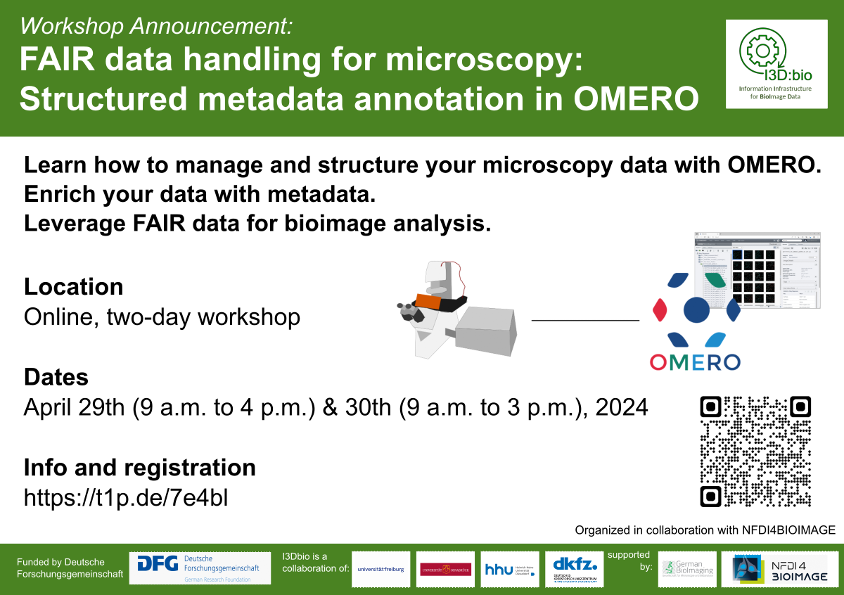
Christian Schmidt

About Christian Schmidt

Contributions by the I3D:bio office are drafted by Christian. Texts are reviewed by members of the I3D:bio team or external collaborators. Feedback welcome.

Fiji-assisted batch analysis using Tags in OMERO

By |2025-08-14T14:48:21+02:00August 11th, 2025|

Fiji-assisted batch analysis using tags in OMERO Let's explore a discrete example! Assumption: We have collected images in OMERO, organized into 6 datasets, with each dataset containing 30 images. The six datasets reflect experiments, each of which contains images from compound-treated biological samples and control samples (solvent only). That means: 10 images [...]

BioImage Analysis and OMERO

By |2025-08-14T08:36:40+02:00August 11th, 2025|

Image Analysis and OMERO While OMERO is not an image analysis software itself, OMERO supports to perform image analysis on the images stored on OMERO's file system through programmatic connections with popular image analysis software or through user-friendly graphical user interfaces. That means, for most data, it is not necessary to download a copy of [...]

Finding and Choosing a Repository

By |2025-08-08T10:59:16+02:00August 1st, 2025|

Back to: Image Data Repositories Finding and Choosing a Data Repository What makes a good repository for imaging data? Public data sharing creates trust in scientific rigor, facilitates reproducibility and enables the potential reuse of data in other projects. Funding agencies may demand that original source data of a publication [...]

Overview of (suitable) storage for bioimaging data

By |2025-07-03T09:52:14+02:00July 2nd, 2025|

What is suitable storage for bioimaging data? Itʼs Not Just About Space - Itʼs About Structure Choosing the right storage solution is crucial - not just for keeping your data safe, but also for ensuring it remains accessible, shareable, and usable for years to come. The choice of storage should therefore be done in [...]

The Specialities of Scientific (Bioimaging) Data Storage

By |2025-07-03T09:56:03+02:00July 2nd, 2025|

What is special about scientific (bioimaging) data storage? Intuitively, most people think of a hard drive (or a USB stick) externally plugged into a computer or internally installed when we talk about storage. While this is sufficient for many everyday cases, scientific-scale data storage comes with challenges that go far beyond what we typically encounter [...]

Why storage matters in bioimaging

By |2025-07-03T09:55:16+02:00July 2nd, 2025|

Why does storage matter in bioimaging? Microscopy experiments create digital images (pixel data) accompanied by accessory data about the image acquisition settings, acquisition date, etc. (metadata). Inside the computer, this information is stored in binary code as a series of 1s and 0s (see also Microscopy File Formats). To wrap all the information contained in [...]

Next-generation file formats for bioimaging

By |2025-07-02T12:43:02+02:00July 2nd, 2025|

What are next-generation file formats (NGFF)? Structure of a classical file format As introduced in Bioimaging File Formats explained, microscopy image data can be written in various different file formats, mostly defined by the vendors of the microscopes used. Most of these files and also the "de facto" open community standard file "OME-TIFF" are [...]

Practical guide to data management around the bioimage data life cycle for core facilities

By |2024-07-29T15:28:31+02:00July 29th, 2024|

Back to: Data Life Cycle Research data management for bioimaging confronts scientists and core facility staff with significant challenges. Orienting along the data life cycle concept for bioimaging data helps to create an overview and define necessary measures at research institutions to advance the data management capabilities. An asset is the implementation of a [...]

Go to Top