On this webpage, we provide an overview on microscopy and image analysis resources, as well as tips & tricks for microscopy. Get in touch with us to add further resources, and let us know if there are questions. We hope you have fun exploring!
A collection of online tutorials, courses, podcasts provided by facilities, the scientific community, and companies.
Microscopy in General
- The microcourses (Harvard University)
- iBiology provides open-access free videos of seminars and short talks given by the world’s leading scientists:
- Online microscopy course “MyScope” by Microscopy Australia: Microscopy Basics & Light Microscopy
- Introduction to Confocal Microscopy by OiVM Baylor College of Medicine
Online teaching by scientific community
- Online FLIM Symposium – Beginners Guide to Fluorescence Lifetime Imaging Microscopy (November 2020)
- Educational Resources by Biopolis Dresden Imaging Platform (BioDIP)
- Janelia’s Optical Interest Group (OIG) playlist
- Optical Microscopy Primer A comprehensive web site at the Florida State University
Online teaching materials by companies
- Abbelight: (recorded) Webinars
- Abberior: Microscopy tutorials on superresolution
- Andor: (recorded) Webinar Expansion Microscopy
- Aurox: recorded lectures of Aurox virtual conference on Microscopy (added 14th April 2020)
- Ibidi: Webinars
- Molecular Probes / Thermo Fisher Scientific: School of Fluorescence
- Mountains: Webinar, Best practices for 3D reconstruction of 2D SEM images
- Nanosurf: Videos and Webinars
- Nikon: MicroscopyU
- Photometrics: Remote learning seminars
- Phasics: webinar series covering Phasics unique wavefront sensing technology and applications for laser, optics, and material metrology
- Zeiss:
- Online microscopy campus for light microscopy: Interactive tutorials and articles on principles
- Microscopy Insights Hub: Discover and share on-demand webinars, how-to videos, and white papers for your field of application.
- Academy Microscopy: Explore our library of learning courses and videos tutorials to select microscopy content that best fits to your needs, instruments and software in our ZEISS Portal and login for free (registration required).
- Microscopist Podcast: Revealing, entertaining, and personal meetings with the great microscopists of our time.
Additional Links
- The Microlist provides a general collection of useful websites and tools.
- Find here a similar list provided by The Royal Microscopy Society (RMS).
- QUAREP-LiMi – Quality Assesment and Reproducibility for Intruments & Images in Light Microscopy – A community with the aim to improve quality assessment and quality control in light microscopy
GerBI-GMB community curated collection of short video tutorials and protocols with helpful tips & tricks for microscopy
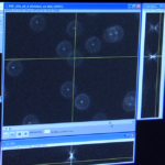
How to measure a Point Spread Function (PSF)
Measuring the point spread function of a microscope is important to determine the lateral and axial resolution capabilities of a microscope. Nico Stuurmann shows how to measure the point spread function of a microscope (iBiology). The video includes the preparation of a bead slide and explains the appropriate image acquisition. A detailed protocol including slide preparation, image acquisition and PSF analysis is provided by QUAREP-LiMi.
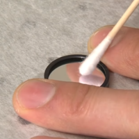
How to clean objective lenses and filters
Cleaning microscope optics like objective lenses and filters is important, because dirt and residues on their surfaces can degrade image quality. Kurt Thorn describes in this iBiology video how to inspect and clean lenses and filters in a way that these expensive components will not be damaged during the process.
 How to find the focal plane
Focusing the specimen is one of the first steps in microscopy. In this iBiology video Ron Vale provides a detailed explanation and helpful tips how to easily find the focal plane without harming your sample or damaging expensive objective lenses.
 How to set up Koehler Illumination
Koehler illumination is the preferred method for obtaining even illumination of the focal plane of the specimen and is a critical alignment procedure in microscopy. Ron Vale shows in this iBiology video a step-by-step procedure of aligning the lamp and condenser to achieve Koehler illumination.
 Live cell imaging and environmental control
To collect scientifically meaningful data from live cell imaging experiments, it is crucial keep the cells on a microscope in a stable and controlled environment. Kurt Thorn explains how to control temperature, humidity, and atmosphere (CO2 concentration) on a microscope. In the iBiology video he discusses simple and more complex systems.
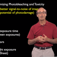 How to minimize photobleaching and phototoxicity
Minimizing phototoxicity during fluorescent microscopy increases the lifetime and viability of your sample. Ron Vale illustrates in this iBiology video how illumination of a specimen can cause photobleaching and protein damage and gives us tips about minimizing damage from fluorescence. Ways to minimize such problems include strategies for minimizing light exposure and removing molecular oxygen from in vitro reactions.
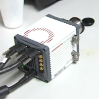 How to calibrate a camera
In this iBiology video about camera calibration, Nico Stuurman shows you how to measure a camera dark image, calculate the readout noise, estimate the dark current, get a camera flat field image using a plastic cup and a cell phone screen, and estimate the photon conversion factor and full well capacity by measuring a photon transfer curve.
A collection of online tutorials, courses, podcasts provided by facilities, the scientific community, and companies.
Community Support by Software Companies
- SVI: Huygens trial license
- Imaris: https://imaris.oxinst.com/imaris-viewer
- OxfordInstruments: Imaris satellite licences
- SVI: Web based deconvolution freely available via SVI-HRM server
- Zeiss: ZEN free offline license 90 days
Online Teaching Materials by Scientific Community
- NEUBIAS Image Analysis Training Resources https://neubias.github.io/training-resources/index.html
- Robert Haase’s “Applied Bio-Image Analysis lecture” at Biotec Dresden on Youtube
- DataViz Academy: A collection of data visualization talks for biomedical scientists. Organized by the MSNZ of the TU Dresden.
- Dave Mason’s Fiji online course
- QuPath, an open powerful, software platform for whole slide image analysis, Intro in their youtube channel
- Pete Bankhead’s open Introduction to Image Analysis book
- iBiology bioimage analysis course: https://www.ibiology.org/online-biology-courses/bioimage-analysis-course/
- Community thread about bioimage analysis training material
https://forum.image.sc/t/bioimage-analysis-recommended-reading-and-viewing/28051
Online Teaching Materials by Companies
- SVI: Deconvolution etc. Youtube channel
- Imaris: recorded Imaris Homeschool webinar series
- Mountains: Webinar, Best practices for 3D reconstruction of 2D SEM images
- Zeiss: Zen knowledge base with tutorials for image analysis and processing. ZEISS Experts walk you through image acquisition as well as image analysis workflows and tutorials for python scripting (from basic to advanced in 72 video tutorials).
Additional Links Related to Image Analysis
- The Microlist provides a general collection of useful websites and tools.
- Find here a similar list provided by The Royal Microscopy Society (RMS).
- QUAREP-LiMi – Quality Assesment and Reproducibility for Intruments & Images in Light Microscopy – A community with the aim to improve quality assessment and quality control in light microscopy
Collection of resources provided via Microscopy DB
You can subsample the results by using the filter option
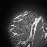
Header image: Drosophila melanogaster egg chamber preparation. Actin cytoskeleton with depth coding. Olympus FV3000, Thomas Zobel, CAi HHU